Timeline of brain cancer
The content on this page is forked from the English Wikipedia page entitled "Timeline of brain cancer". The original page still exists at Timeline of brain cancer. The original content was released under the Creative Commons Attribution/Share-Alike License (CC-BY-SA), so this page inherits this license.
This is a timeline of brain cancer, describing especially major discoveries, advances in treatment and major organizations.
Contents
Big picture
| Year/period | Key developments |
|---|---|
| 19th century | First surgery for a brain tumor is performed in this century.[1] |
| 1920s | Electroencephalograms (EEG) in humans develop as a method to record activity in the brain.[2] |
| 1970s | Computed tomography (CT) scanning is developed and provides first clear image of brain tumors. First promising chemotherapy for glioma is developed. Radiation is established as standard treatment for glioblastoma.[3] |
| 1980s | Magnetic resonance imaging is introduced and gains widespread use. It eventually replaces CT scanning as the primary imaging tool for brain tumors. Gamma knife therapy is introduced for treating brain tumors.[3] |
| 1990s | Advances in chemotherapy and radiation increases survival for patients. Classification for brain tumors unifies into one universal system.[3] |
| 2000s | The Cancer Genome Atlas project launches.[3] |
| 2010s | Today, cancer immunotherapy is being actively studied.[4] Primary brain tumors occur in around 250,000 people a year globally, making up less than 2% of cancers.[5] Figures for incidence on brain cancer show predominance in more developed countries.[6] |
Numerical and visual data
Google Scholar
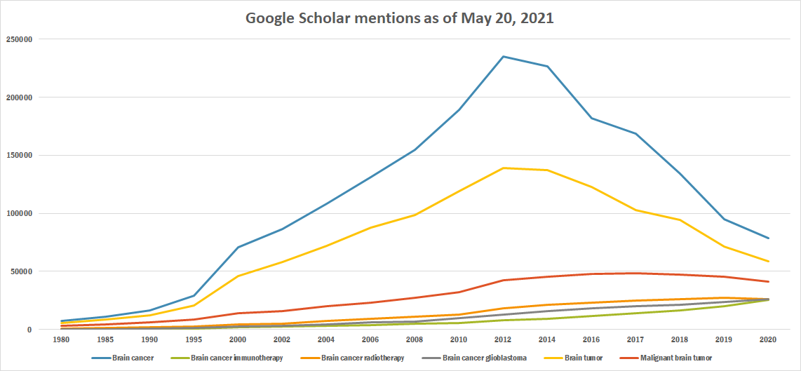
The following table summarizes per-year mentions on Google Scholar as of May 20, 2021.
| Year | Brain cancer | Brain cancer immunotherapy | Brain cancer radiotherapy | Brain cancer glioblastoma | Brain tumor | Malignant brain tumor |
|---|---|---|---|---|---|---|
| 1980 | 7,680 | 285 | 883 | 284 | 5,860 | 3,010 |
| 1985 | 11,000 | 285 | 1,290 | 435 | 8,810 | 4,480 |
| 1990 | 16,200 | 511 | 1,830 | 807 | 12,500 | 6,160 |
| 1995 | 29,400 | 831 | 2,640 | 1,540 | 20,400 | 8,340 |
| 2000 | 70,900 | 1,910 | 4,390 | 2,820 | 45,900 | 13,900 |
| 2002 | 86,500 | 2,300 | 5,220 | 3,220 | 57,900 | 16,000 |
| 2004 | 108,000 | 3,210 | 7,390 | 4,230 | 72,100 | 19,800 |
| 2006 | 131,000 | 3,830 | 9,200 | 5,920 | 87,500 | 23,200 |
| 2008 | 155,000 | 4,950 | 11,300 | 6,800 | 98,700 | 27,400 |
| 2010 | 189,000 | 5,850 | 13,100 | 9,560 | 119,000 | 32,200 |
| 2012 | 235,000 | 7,700 | 18,200 | 13,100 | 139,000 | 42,200 |
| 2014 | 227,000 | 9,340 | 21,100 | 15,800 | 137,000 | 45,700 |
| 2016 | 182,000 | 11,900 | 23,200 | 18,000 | 123,000 | 47,700 |
| 2017 | 169,000 | 14,200 | 24,900 | 19,900 | 103,000 | 48,500 |
| 2018 | 134,000 | 16,400 | 26,400 | 21,500 | 94,200 | 47,100 |
| 2019 | 94,900 | 19,800 | 27,200 | 23,700 | 71,600 | 45,500 |
| 2020 | 78,500 | 25,400 | 25,900 | 26,100 | 58,500 | 41,100 |
Google Trends
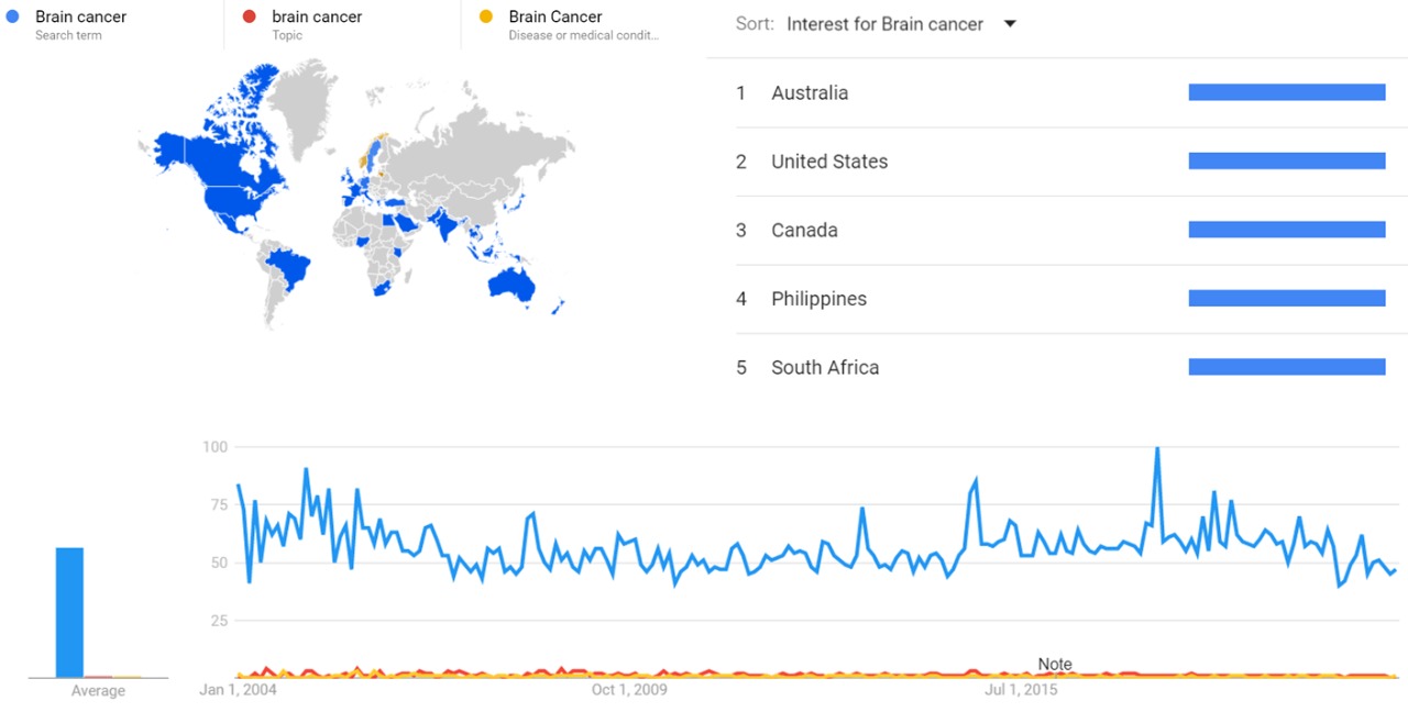
The comparative chart below shows Google Trends data Brain cancer (search term, topic and Disease or medical condition) from January 2004 to January 2021, when the screenshot was taken.[7]
Google Ngram Viewer
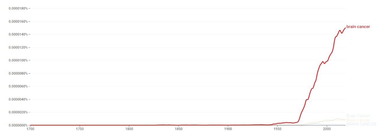
The chart below shows Google Ngram Viewer data for Brain cancer from 1700 to 2019.[8]
Wikipedia views
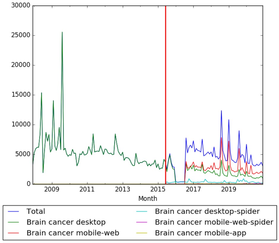
The chart below shows Wikipedia views of the article brain cancer on desktop from December 2007, and on mobile-web, desktop-spider, mobile-web-spider and mobile app, from June 2015; to January 2021.
Full timeline
| Year/period | Type of event | Event | Location |
|---|---|---|---|
| 1879 | Achievement | Scottish surgeon William Macewen performs the first successful brain tumor removal in a young woman.[1] | |
| 1904 | Development | Medulloepithelioma (a rare brain tumor thought to stem from cells of the embryonic medullary cavity) is first described by Frederick Verhoeff.[9] | |
| 1907 | Development | First description of the clinical syndrome of neuroblastoma of the adrenals by English surgeon Jonathan Hutchinson. However, the pathologic interpretation of the disease is not established until several years later.[10] | |
| 1908 | Development | Subependymal giant cell astrocytoma (a low-grade astrocytic brain tumor) is first described.[11] | |
| 1918 | Development | Pilocytic astrocytoma (a brain tumor that occurs more often in children and young adults) is first described.[12] | |
| 1920 | Development | Lhermitte–Duclos disease (rare tumor of the cerebellum) is first described by French neurologist Jacques Jean Lhermitte and P. Duclos.[13] | |
| 1925 | Development | Medulloblastoma (the most common type of pediatric malignant primary brain tumor) is first described.[14] | |
| 1926 | Development | Percival Bailey and Harvey Cushing introduce the term glioblastoma multiforme, based on the idea that the tumor originates from primitive precursors of glial cells (glioblasts), and the highly variable appearance due to the presence of necrosis, hemorrhage and cysts (multiform).[15] | |
| 1930 | Development | Astroblastoma, a rare glial tumor, is first described.[16] | |
| 1936 | Development | American neurophysiologist William Grey Walter first identifies the association between localized slow waves on electroencephalograms and tumors of the cerebral hemispheres. The term "delta waves" is introduced by Walter.[2] | |
| 1936 | Development | Optic nerve sheath meningioma (a rare benign tumor of the optic nerve), is first described.[17] | |
| 1938 | Gliomatosis cerebri (a rare primary brain tumor) is first described.[18] | ||
| 1942 | Development | Hemangiopericytoma (a tumor located in the cerebral cavity) is first characterized.[19] | |
| 1945 | Development | Subependymoma (a rare form of ependymal tumor) is first described.[20] | |
| 1955 | Development | Choroid plexus papilloma (a rare benign neuroepithelial intraventricular WHO grade I lesion found in the choroid plexus) is first reported.[21] | |
| 1958 | Treatment | Dexamethasone is first synthesized. To date it remains the most favorable drug for brain cancer patients.[22] | |
| 1971 | Development | Trilateral retinoblastoma (a malignant midline primitive neuroectodermal tumor occurring in patients with retinoblastoma) is first described.[23] | |
| 1971 | Development | British company EMI develops CT scanner that provides pictures of patients brains to be seen for the first time.[24] | UK |
| 1973 | Organization | American Brain Tumor Association is founded as a nonprofit organization. It provides support services and programs to brain tumor patients and their families, and funds brain tumor research.[25] | Chicago, Illinois, US |
| 1975 | Treatment | Radiation therapy becomes standard treatment for glioblastoma, showing it extends median survival from 3 months to about 9 months in patients.[3] | |
| 1978 | Development | Researchers at EMI Laboratories obtain the first Magnetic resonance imaging (MR) of a human brain.[26] | UK |
| 1979 | Development | Pleomorphic Xanthoastrocytoma (a rare tumor located throughout the supratentorial compartment in the brain) is first described.[16] | |
| 1982 | Development | Central neurocytoma (an extremely rare, ordinarily benign intraventricular brain tumor), is first described.[27] | |
| 1988 | Organization | The Children's Brain Tumor Foundation is established. It funds research in brain cancer.[28][29] | New York City, US |
| 1993 | Discovery | Large analysis shows that adding chemotherapy to radiation therapy helps patients with surgically treated malignant gliomas live longer compared to radiation therapy alone.[3] | |
| 1993 | Development | World Health Organization develops universal system for classifying brain tumors with aims at improving communication and translate research findings.[3] | |
| 1994 | Development | The first study of the human brain at 3.0 T is published.[30] | |
| 1994 | Development | US National Cancer Institute establishes brain tumor research networks for adults and children, comprising brain cancer experts from academic centers who collaborate to evaluate novel therapies against brain cancer.[3] | United States |
| 1996 | Discovery | Researchers at University of Washington find that brain cancer is related to low levels of protein MGMT.[31] | United States |
| 1996 | Development | Giant cell ependymoma (an uncommon neuroepithelial tumor located in the central nervous system) is first described.[32] | |
| 1997 | Development | Researchers at Washington University first develop susceptibility weighted imaging, which can help determine the status of a tumor in the brain.[33] | US |
| 1997 | Treatment | British doctors make use of first laser system to treat brain tumors by destroying cancerous tissue.[34] | UK |
| 1998 | Development | The first study of the human brain at 8 T is published.[35] | |
| 1998 | Organization | The German Brain Tumor Association (Deutsche Hirntumorhilfe) is founded. It supports research especially in the field of neurooncology.[36] | Leipzig, Germany |
| 2001 | Achievement | First successful application of retroviral replicating vectors towards transducing cell lines derived from solid tumors.[37] | |
| 2001 | Organization | Accelerate Brain Cancer Cure is founded as a nonprofit organization. It awards grants to leading brain cancer researchers and partners with the medical, academic, business, and government sectors.[38][39] | Washington, DC., US |
| 2002 | Epidemiology | Study reports that brain tumors represent about 1–2 percent of all newly diagnosed tumors, and accounts for about 2 percent of all cancer–related deaths.[40] | |
| 2003 | Treatment | Use of chemotherapy wafer containing carmustine (BCNU) is found to delay tumor growth and improve overall survival in some patients with gliomas.[3] | |
| 2004 | Treatment | International study shows that giving low doses of temozolomide at the start of the treatment proves very effective against brain cancer.[41][42] | |
| 2005 | Discovery | Researchers discover that patients with tumors carrying a specific alteration in a gene MGMT benefit from temozolomide (Temodar) therapy.[3] | |
| 2005 | Study | US National Cancer Institute and US National Genome Research Institute launch The Cancer Genome Atlas Project, with the goal of mapping the genetic changes involved in glioblastoma and other cancers.[3] | United States |
| 2005 | Development | Angiocentric Glioma (a tumor of the central nervous system), is described.[16] | |
| 2006 | Discovery | Researchers discover distinct subtypes of astrocytoma tumors, each one with unique biological features that appear to influence the tumor's behavior and response to certain therapies.[3][43] | |
| 2008 | Discovery | Researchers find that family members of patients with brain cancer, particularly astrocytoma, may inherit a higher risk of the same diagnosis.[44] | |
| 2008 | Discovery | The Cancer Genome Atlas Project reports the identification of several key mutations in the ERBB2, NF1 and TP53 genes, that are involved in triggering the development and spread of glioblastoma.[3] | United States |
| 2008 | Treatment | FDA approves bevacizumab to treat glioblastoma.[3] | United States |
| 2009 | Discovery | Researchers discover that tumors with an alteration in the IDH1 or IDH2 genes are less aggressive than those without the mutation. This finding would eventually enable some patients to safely undergo less intense therapy.[3][45] | |
| 2010 | Discovery | Set of nine genes is found to predict the likelihood that a glioblastoma will respond to therapy.[3] | |
| 2013 | Development | Researchers develop new technique to distinguish tumors from healthy tissue in the brain, using raman scattering microscopy.[46] | US |
| 2016 | Discovery | Study finds that people with higher levels of education may be more likely to develop certain types of brain tumors.[47] | Sweden |
See also
References
- ↑ 1.0 1.1 "History of brain tumor surgery". Retrieved 29 August 2016.
- ↑ 2.0 2.1 "EEG in Brain Tumors". Retrieved 12 September 2016.
- ↑ 3.00 3.01 3.02 3.03 3.04 3.05 3.06 3.07 3.08 3.09 3.10 3.11 3.12 3.13 3.14 3.15 "Cancer Progress". Retrieved 30 August 2016.
- ↑ Bloch, O (2015). "Immunotherapy for malignant gliomas.". Cancer Treatment and Research. 163: 143–58. PMID 25468230. doi:10.1007/978-3-319-12048-5_9.
- ↑ World Cancer Report 2014. World Health Organization. 2014. pp. Chapter 5.16. ISBN 9283204298.
- ↑ Bondy ML, Scheurer ME, Malmer B, et al. (2008). "Brain Tumor Epidemiology: Consensus from the Brain Tumor Epidemiology Consortium (BTEC)". Cancer. 113 (7 Suppl): 1953–1968. PMC 2861559
 . PMID 18798534. doi:10.1002/cncr.23741.
- ↑ "Brain cancer". trends.google.com. Retrieved 13 January 2021.
- ↑ "Brain cancer". books.google.com. Retrieved 15 January 2021.
- ↑ "Intraocular Medulloepithelioma". Archives of Pathology & Laboratory Medicine. 136: 212–216. doi:10.5858/arpa.2010-0669-RS. Retrieved 12 September 2016.
- ↑ "Ocular Complications in Neuroblastoma". American Journal of Ophthalmology. 18: 938–943. doi:10.1016/S0002-9394(35)92482-5.
- ↑ Tahiri Elousrouti L, Lamchahab M, Bougtoub N, Elfatemi H, Chbani L, Harmouch T, Maaroufi M, Amarti Riffi A (2016). "Subependymal giant cell astrocytoma (SEGA): a case report and review of the literature". J Med Case Rep. 10: 35. PMC 4748639
 . PMID 26861567. doi:10.1186/s13256-016-0818-6.
- ↑ "The lost art of localization: Franc Ingraham's legacy in pediatric neurosurgery". Retrieved 12 September 2016.
- ↑ Lhermitte J, Duclos P (1920). "Sur un ganglioneurome diffuse du cortex du cervelet". Bulletin de l'Association Francaise pour l'etude du Cancer. Paris. 9: 99–107.
- ↑ Muzumdar D, Deshpande A, Kumar R, Sharma A, Goel N, Dange N, Shah A, Goel A (2011). "Medulloblastoma in childhood-King Edward Memorial hospital surgical experience and review: Comparative analysis of the case series of 365 patients". J Pediatr Neurosci. 6: S78–85. PMC 3208927
 . PMID 22069434. doi:10.4103/1817-1745.85717.
- ↑ Bailey & Cushing: Tumors of the Glioma Group JB Lippincott, Philadelphia, 1926.Template:Fix/category[page needed]
- ↑ 16.0 16.1 16.2 "Unusual Gliomas". Retrieved 12 September 2016.
- ↑ "Update on Treatment Modalities for Optic Nerve Sheath Meningiomas". Retrieved 12 September 2016.
- ↑ "Gliomatosis Cerebri in a Child: Another Case". Retrieved 12 September 2016.
- ↑ Stout AP, Murray MR (1942). "Hemangiopericytoma: a vascular tumor featuring Zimmermann's pericytes". Ann Surg. 116: 26–33. doi:10.1097/00000658-194207000-00004.
- ↑ "Subependymoma". Retrieved 12 September 2016.
- ↑ Maimone G, Ganau M, Nicassio N, Paterniti S (2013). "Paratrigonal choroid plexus papilloma presenting with satellite multiple supra- and infratentorial hemorrhages. Neuroanatomical basis and pathological hypothesis". Int J Surg Case Rep. 4: 239–42. PMC 3604672
 . PMID 23333804. doi:10.1016/j.ijscr.2012.11.023.
- ↑ Dietrich J, Rao K, Pastorino S, Kesari S (2011). "Corticosteroids in brain cancer patients: benefits and pitfalls". Expert Rev Clin Pharmacol. 4: 233–42. PMC 3109638
 . PMID 21666852. doi:10.1586/ecp.11.1.
- ↑ "Molecular Genetics of Supratentorial Primitive Neuroectodermal Tumors and Pineoblastoma". Retrieved 12 September 2016.
- ↑ "EMI CT brain scanner, England, 1970-1971". Retrieved 29 August 2016.
- ↑ "American Brain Tumor Association". Retrieved 31 August 2016.
- ↑ "Britain's brains produce first NMR scans". New Scientist: 588. 1978.
- ↑ Hassoun, J.; Gambarelli, D.; Grisoli, F.; Pellet, W.; Salamon, G.; Pellissier, JF.; Toga, M. (1982). "Central neurocytoma. An electron-microscopic study of two cases.". Acta Neuropathol. 56 (2): 151–6. PMID 7064664. doi:10.1007/bf00690587.
- ↑ "About Children's Brain Tumor Foundation". Retrieved 31 August 2016.
- ↑ "Children's Brain Tumor Foundation". Retrieved 31 August 2016.
- ↑ Mansfield P, Coxon R, Glover P (May 1994). "Echo-planar imaging of the brain at 3.0: first normal volunteer results.". J Comput Assist Tomogr. 18 (3): 339–43. PMID 8188896. doi:10.1097/00004728-199405000-00001.
- ↑ "Brain cancer, protein levels linked". Retrieved 30 August 2016.
- ↑ Barbagallo GM, Caltabiano R, Parisi G, Albanese V, Lanzafame S (2009). "Giant cell ependymoma of the cervical spinal cord: case report and review of the literature". Eur Spine J. 18 Suppl 2: 186–90. PMC 2899556
 . PMID 18820954. doi:10.1007/s00586-008-0789-4.
- ↑ Reichenbach JR, Venkatesan R, Schillinger DJ, Kido DK, and Haacke EM (1997). "Small vessels in the human brain: MR venography with deoxyhemoglobin as an intrinsic contrast agent". Radiology. 204 (1): 272–277. PMID 9205259. doi:10.1148/radiology.204.1.9205259.
- ↑ "Laser gun surgery for brain cancer". New Straits Times. Retrieved 30 August 2016.
- ↑ Robitaille PM, Abduljalil AM, Kangarlu A, Zhang X, Yu Y, Burgess R, Bair S, Noa P, Yang L, Zhu H, Palmer B, Jiang Z, Chakeres DM, Spigos D (Oct 1998). "Human magnetic resonance imaging at 8 T.". NMR Biomed. 11 (6): 263–5. PMID 9802467. doi:10.1002/(SICI)1099-1492(199810)11:6<263::AID-NBM549>3.0.CO;2-0.
- ↑ "Deutsche Hirntumorhilfe". Retrieved 31 August 2016.
- ↑ Logg CR, Tai CK, Logg A, Anderson WF, Kasahara N (20 May 2001). "A uniquely stable replication-competent retrovirus vector achieves efficient gene delivery in vitro and in solid tumors". Human Gene Therapy. 12 (8): 921–932. PMID 11387057. doi:10.1089/104303401750195881.
- ↑ "Accelerate Brain Cancer Cure". Retrieved 30 August 2016.
- ↑ "Accelerate Brain Cancer Cure". Retrieved 30 August 2016.
- ↑ Ekman, M.; Westphal, M. "Cost of brain tumor in Europe".
- ↑ Gerber DE, Grossman SA, Zeltzman M, Parisi MA, Kleinberg L (2007). "The impact of thrombocytopenia from temozolomide and radiation in newly diagnosed adults with high-grade gliomas". Neuro-oncology. 9: 47–52. PMC 1828105
 . PMID 17108062. doi:10.1215/15228517-2006-024.
- ↑ "Early drug treatment is effective against brain cancer". The Day. Retrieved 30 August 2016.
- ↑ "Molecular subclasses of high-grade glioma predict prognosis, delineate a pattern of disease progression, and resemble stages in neurogenesis". Cancer Cell. 9: 157–173. doi:10.1016/j.ccr.2006.02.019.
- ↑ "Brain Cancer in the Family Raises Risk". Retrieved 29 August 2016.
- ↑ Yan H, Parsons DW, Jin G, et al. (February 2009). "IDH1 and IDH2 Mutations in Gliomas". New England Journal of Medicine. 360: 765–773. PMC 2820383
 . PMID 19228619. doi:10.1056/NEJMoa0808710.
- ↑ "Laser imaging spots brain cancer". Retrieved 30 August 2016.
- ↑ "Brain Tumor Risk Linked with Higher Education, Study Finds". Retrieved 29 August 2016.


