Timeline of radiology
This is a tiemeline of radiology, listing important events in the development of the field.
Big picture
| Time period | Development summary |
|---|---|
| 19th century | Late in the century, Wilhelm Röntgen first discovers the X-ray. |
| 20th century | Soon after the turn of the century, lay x-ray operators start being appointed as assistants.[1] In the 1950s comes the development of image intensifier and x-ray television.[2] In the 1960s, ultrasound gains popularity.[2] The 1970s are known as the "golden decade" of radiology, when the CT scanner opens up new opportunities and discoveries which would be further developed in the following decades.[3] Magnetic resonance imaging develops.[2] In the 1980s, position emission tomography (PET) emerges as new technology.[1] Clinical MRI is also introduced in the 1980s.[4] in the 1990s, a rising interest in the construction of a combined PET-CT scanner emerges.[5] |
| 21st century | PET/CT becomes one of the fastest growing medical imaging modalities, rivaling the growth of MR during the 1980s and 1990s.[6] As of 2008, over 2500 PET-CT scanners are operational worldwide.[6] |
Full timeline
| Year | Event type | Details | Country/region |
|---|---|---|---|
| 1895 | Field development | German physicist Wilhelm Röntgen first discovers the X-ray.[2][7] | |
| 1896 | Field development | French physicist Antoine-Henri Becquerel discovers radioactivity.[7] | France |
| 1896 | Literature | Journal Archives of Clinical Skiagraphy launches as the first radiology scientific journal.[7][8] | United Kingdom |
| 1896 | Field development | After learning about Röntgen’s discoveries, American inventor Thomas Edison invents fluoroscopy. Fluoroscopic screens would be then used as an alternation to still x-ray images for some time.[9][7] | United States |
| 1898 | Literature | Marie Curie publishes her paper Rays emitted by uranium and thorium compounds.[7] | France |
| 1900 | Organization | The American Roentgen Ray Society (ARRS) is founded.[1] | United States |
| 1901 | Award | Wilhelm Röntgen is awarded the Nobel Prize in Physics for his contribution to the study of radiation.[10] | |
| 1903 | Field development | Lay x-ray operators start being appointed as assistants.[1] | |
| 1913 | Field development | German surgeon Albert Salomon initiates research leading to mammography.[7] Solomon becomes the first to use x-ray imaging to view the gross anatomy of mastectomy specimens and is the first to demonstrate successful visualization of microcalcifications.[11][12][13] | Germany |
| 1914 – 1918 | Field development | Radiological equipment is used in field hospitals during World War I.[1] | |
| 1915 | Organization | The Western Roentgen Society is founded in Chicago.[14][15][16] | United States |
| 1916 | Organization | The Russian Society of Radiology is founded. It is the oldest and the only national society of Russian radiologists, nuclear medicine specialists, medical physics, radiographers, technitians and other specialists related to radiology, diagnostic and interventional imaging.[17] | Russia |
| 1918 | Field development | George Eastman introduces film, which would replace radiographs made onto glass photographic plates.[2][9] | |
| 1918 | Literature | The Radiological Society of North America publishes journal Radiology.[14] | United States |
| 1920 | Organization | The Society of Radiographers is formed in the United Kingdom as a trade union and professional body for x-ray and radiation technicians.[2][9] | United Kingdom |
| 1920 | Organization | The American Society of Radiologic Technologists is founded.[18] | United States |
| 1921 | Field development | Diagnostic radiology takes a great leap forward with the introduction of pneumoventriculography and pneumoencephalography.[19] | |
| 1923 | Literature | Monthly, peer reviewed, medical journal Radiology is released by the Radiological Society of North America.[20] | United States |
| 1927 | Field development | Portuguese neurologist António Egas Moniz develops cerebral angiography, a technique using X-rays to visualize arteries and veins that are transiently opacified with the injection of high density agent.[21][22][23][7] | Portugal |
| 1934 | Field development | French scientists Frederic and Irène Joliot-Curie artificially produce radioisotopes.[7] | France |
| 1935 | The higher radiological qualification known as the Fellowship is created by The British Association of Radiologists.[1] | United Kingdom | |
| 1935 | Organization | The Society of Radiotherapists of Great Britain and Ireland is established.[1] | United Kingdom |
| 1936 | Field development | American hematologist John H. Lawrence of the University of California, Berkeley introduces phosphorus-32 for the treatment of leukemia.[24][25][26] | United States |
| 1939 | Literature | Kitty Clark publishes Clark’s Positioning in Radiography, which would become a preeminent text on positioning technique for diagnostic radiographers.[27][28][29][7] | |
| 1939 | Organization | The Faculty of Radiologists is formed, amalgamating the British Association of Radiologists and the Society of Radiotherapists of Great Britain and Ireland.[1] | United Kingdom |
| 1953 | Field development | Swedish radiologist Sven Ivar Seldinger pioneers the Seldinger technique, laying down the foundation of interventional radiology.[30][7] | Sweden |
| 1954 | Field development | David Kuhl, a medical student at the University of Pennsylvania, invents the "photoscan", which would replace the scintiscanner.[31] | |
| 1958 | Field development | Scottish physician Ian Donald develops the first medically used ultrasound to observe the health and growth of fetuses. Donald also uses the ultrasound to study lumps, cysts, and fibroids. Donald, Together with engineer Tom Brown, develop a portable ultrasound machine to be used on patients.[9][7] | United Kingdom |
| 1961 | Field development | James Robertson, working at the Brookhaven National Laboratory, builds the first single-plane positron emission tomography (PET) scan.[9] | United States |
| 1962 | Field development | American scientist David E. Kuhl introduces emission reconstruction tomography. This method later becomes known as SPECT and PET.[1] | United States |
| 1962 | Organization | The European Association of Radiology is established.[32][33][34] | |
| 1963 | Field development | American radiologist Charles Theodore Dotter first proposes the idea of interventional radiology.[30] | United States |
| 1964 | Field development | Charles Theodore Dotter introduces image-guided intervention.[7] | |
| 1965 | Field development | Transcatheter arterial embolization becomes one of the most important basic techniques for interventional radiology.[30] | |
| 1965 | Literature | Benjamin Felson publishes his Principles of Chest Roentgenology.[35][36][7] | |
| 1967 | Field development | The first clinical use of magnetic resonance imaging takes place in England.[1] | United Kingdom |
| 1967 | Field development | Transjugular intrahepatic portosystemic stent-shunt becomes a comprehensive interventional radiology technology, in which the biliary system can be reached through a jugular vein.[30] | |
| 1971 | Field development | English electrical engineer Godfrey Hounsfield builds the prototype computerized tomography (CT) machine, which utilizes both x-rays and computer software to create cross-sectional images of the body. In the same year, the first successful medical scan using this machine is done on a live patient.[9] | United Kingdom |
| 1972 | Field development | Godfrey Hounsfield introduces the first clinical prototype of CT scanner.[4][7][2][5][37][38] | United Kingdom |
| 1972 | Field development | The EMI parallel beam scanner is introduced.[37] | |
| 1972 | Field development | Non-vascular interventional techniques becomes an important branch of interventional radiology.[30] | |
| 1973 | Field development | American chemist Paul Lauterbur develops the way to generate the first two-dimensional and three-dimensional magnetic resonance images (MRIs). In the same year, Lauterbur publishes the first nuclear magnetic resonance image.[9][4] | United States |
| 1975 | Field development | Frank T Farmer gives an interesting historical review of the physical basis of radiology and demonstrates diffraction patterns as obtained by Von Laue.[39] | |
| 1975 | Field development | Michael E. Phelps, Michel Ter-Pogossian, and co-workers at Washington University School of Medicine introduce the modern PET scanner. The design is a ring system surounding the patient.[5] | United States |
| 1975 – 1980 | Field development | "Real-time" ultrasound machines are introduced.[2] | |
| 1977 | Field development | English physicist Peter Mansfield of the University of Nottingham describes the general principles of echo-planar imaging.[40] Mansfield develops echo-planar imaging for MRIs by mathematically analyzing the radio signals from magnetic resonance imaging. This development allows for images to be collected much faster than previously possible.[9] | United Kingdom |
| 1977 | Field development | American physician Raymond Damadian completes the first MRI (magnetic resonance imaging).[41][42][43] | United States |
| 1979 | Award | South African physicist Allan McLeod Cormack and Godfrey Hounsfield are awarded the Nobel Prize in Physiology or Medicine "for the development of computer assisted tomography". | |
| c.1980 | Field development | The first commercial PET scanner is introduced.[5] | |
| 1985 | Field development | Argentine physician Julio Palmaz develops the balloon-expandable stent, thus transforming interventional radiology.[44][45][46][9] | United States |
| 1989 | Field development | 3D data acquisition becomes available with the introduction of spiral CT by W.A. Kalender.[37][47][48] | |
| 1991 | Field development | The first functional MRI (fMRI) of the brain is conducted by Belliveau et al.[38] | |
| 1998 | Field development | Ronald Nutt and David Townsend present the first combined PET-CT prototype scanner. It combines positron emission tomography and computerized tomography in such a way as to make it easier for physicians to locate tumors and other structures on the images. The combination also makes it much easier and less expensive for physicians and hospitals to have access to both forms of technology.[9][5] | United States |
| 2000 | The PET-CT scanner, attributed to David Townsend and Ronald Nutt, is named by TIME Magazine as the medical invention of the year.[5] | ||
| 2001 | Field development | The first commercial PET-CT system, Discovery LS, is released by American multinational GE Healthcare. It consists in a single-slice spiral CT integrated with a PET scanner with BGO detectors.[5] | United States |
| 2003 | Award | Peter Mansfield shares with Paul Lauterbur the Nobel Prize in Physiology or Medicine, , for discoveries concerning Magnetic Resonance Imaging (MRI) | |
| 2004 | Field development | The Siemens 64-slice spiral CT is introduced.[37] | |
| 2006 | Field development | PET-only scanners are no longer obtainable as major medical centers and clinics opt for PET/CT to replace their PET-only scanners and newly-established diagnostic imaging centers go directly to PET-CT.[6] | |
| 2008 | Field development | As of date, over 2500 PET-CT scanners are operational worldwide.[6] | |
| 2012 | The International Day of Radiology (IDoR) is introduced. It is celebrated on November 8 each year.[7] |
Numerical and visual data
Google Scholar
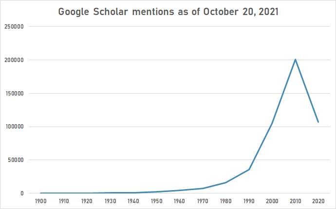
The following table summarizes per-year mentions on Google Scholar as of October 20, 2021.
| Year | radiology |
|---|---|
| 1900 | 119 |
| 1910 | 352 |
| 1920 | 501 |
| 1930 | 980 |
| 1940 | 1,260 |
| 1950 | 2,250 |
| 1960 | 4,270 |
| 1970 | 7,440 |
| 1980 | 16,500 |
| 1990 | 36,000 |
| 2000 | 105,000 |
| 2010 | 201,000 |
| 2020 | 107,000 |

Google Trends
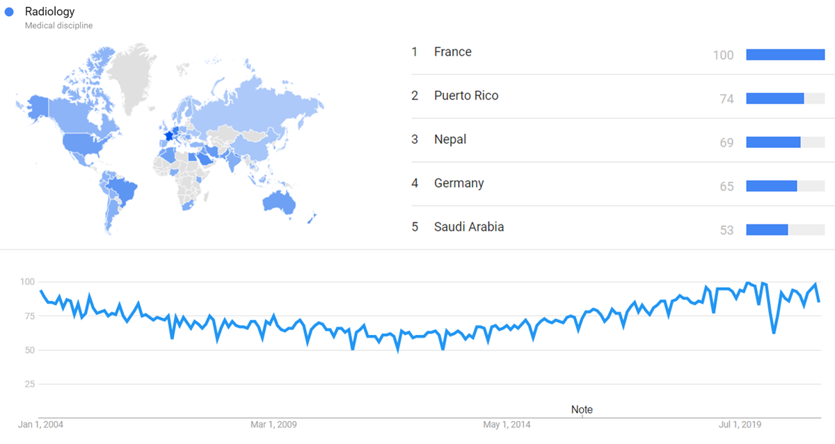
The chart below shows Google Trends data for Radiology (Medical discipline), from January 2004 to April 2021, when the screenshot was taken. Interest is also ranked by country and displayed on world map.[49]

Google Ngram Viewer
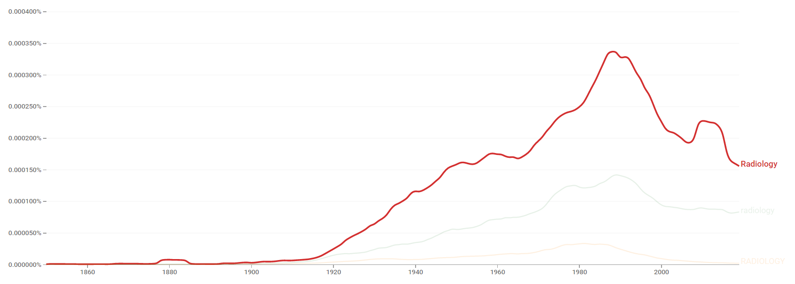
The chart below shows Google Ngram Viewer data for Radiology, from 1850 to 2019.[50]

Wikipedia Views
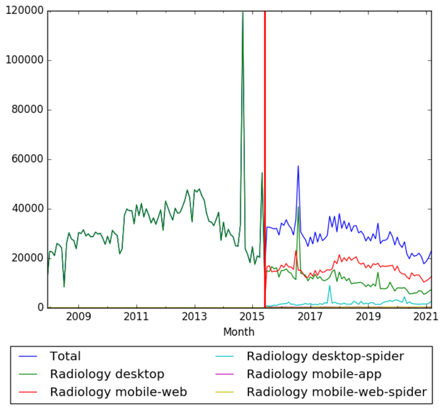
The chart below shows pageviews of the English Wikipedia article Radiology, on desktop from December 2007, and on mobile-web, desktop-spider, mobile-web-spider and mobile app, from July 2015; to March 2021.[51]

Meta information on the timeline
How the timeline was built
The initial version of the timeline was written by User:Sebastian.
Funding information for this timeline is available.
Feedback and comments
Feedback for the timeline can be provided at the following places:
- FIXME
What the timeline is still missing
Timeline update strategy
See also
External links
References
- ↑ 1.00 1.01 1.02 1.03 1.04 1.05 1.06 1.07 1.08 1.09 "Historical timeline". rcr.ac.uk. Retrieved 14 August 2018.
- ↑ 2.0 2.1 2.2 2.3 2.4 2.5 2.6 2.7 "Origins of radiology". bir.org.uk. Retrieved 9 August 2018.
- ↑ "History of radiology". bir.org.uk. Retrieved 9 August 2018.
- ↑ 4.0 4.1 4.2 Orrison, William W.; Lewine, Jeffrey; Sanders, John; Hartshorne, Michael F. Functional Brain Imaging.
- ↑ 5.0 5.1 5.2 5.3 5.4 5.5 5.6 Functional Imaging in Oncology: Biophysical Basis and Technical Approaches -, Volume 1 (Antonio Luna, Joan C. Vilanova, L. Celso Hygino da Cruz Jr., Santiago E. Rossi ed.).
- ↑ 6.0 6.1 6.2 6.3 Townsend, David W. "Combined PET/CT: the historical perspective". doi:10.1053/j.sult.2008.05.006. PMID 18795489.
{{cite journal}}: Cite journal requires|journal=(help) - ↑ 7.00 7.01 7.02 7.03 7.04 7.05 7.06 7.07 7.08 7.09 7.10 7.11 7.12 7.13 7.14 "History of radiology". radiopaedia.org. Retrieved 13 August 2018.
- ↑ "Archives of Clinical Skiagraphy". radiopaedia.org. Retrieved 13 August 2018.
- ↑ 9.00 9.01 9.02 9.03 9.04 9.05 9.06 9.07 9.08 9.09 "10-Minute History of Radiology: Overview of Monumental Inventions". bicrad.com. Retrieved 13 August 2018.
- ↑ "An Illuminating Accident". nobelprize.org. Retrieved 13 September 2018.
- ↑ Bassett, Lawrence W.; Mahoney, Mary C; Apple, Sophia; D'Orsi, Carl. Breast Imaging Expert Radiology Series E-Book.
- ↑ Kaiser, Werner A. Signs in MR-Mammography.
- ↑ Classic Papers in Modern Diagnostic Radiology (Adrian M. K. Thomas, Arpan K. Banerjee, Uwe Busch ed.).
- ↑ 14.0 14.1 Adler, Arlene M.; Carlton, Richard R. Introduction to Radiologic and Imaging Sciences and Patient Care - E-Book.
- ↑ Mould, R.F. A Century of X-Rays and Radioactivity in Medicine: With Emphasis on Photographic Records of the Early Years.
- ↑ Hussey, David H.; Beck, Bill. ASTRO: A Celebration of 50 Years.
- ↑ "Russian Society of Radiology". healthmanagement.org. Retrieved 17 November 2018.
- ↑ "History of the American Society of Radiologic Technologists". asrt.org. Retrieved 13 August 2018.
- ↑ Tandon, Prakash Narain; Ramamurthi, Ravi. Textbook of Neurosurgery, Third Edition, Three Volume Set.
- ↑ "Medical Journals Recommended by Neil Gerardo". mrx.com. Retrieved 29 August 2018.
- ↑ The Medical Basis of Psychiatry (S. Hossein Fatemi, Paula J. Clayton ed.).
- ↑ Oakes, Elizabeth H. Encyclopedia of World Scientists.
- ↑ Neuroimaging, Part 1.
- ↑ Marks, Geoffrey; Beatty, William K. The Precious Metals of Medicine.
- ↑ Oreskes, Naomi; Krige, John. Science and Technology in the Global Cold War.
- ↑ Positron Emission Tomography: Basic Sciences (Dale L. Bailey, David W. Townsend, Peter E. Valk, Michael N. Maisey ed.).
- ↑ "Clark's Positioning in Radiography 13E". crcpress.com. Retrieved 14 September 2018.
- ↑ "CLARK'S POSITIONING RADIOGRAPHY 12th Edition Hardcover – 26 Aug 2005". amazon.co.uk. Retrieved 14 September 2018.
- ↑ Yeung, Andy WK. "Tube shift or tube tilt? The terminology of dental radiography is heterogeneous relative to radiological convention". hkmj.org. Retrieved 14 September 2018.
- ↑ 30.0 30.1 30.2 30.3 30.4 Tang, Z; Jia, A; Li, L; Li, C. "[Brief history of interventional radiology]". PMID 25208839.
{{cite journal}}: Cite journal requires|journal=(help) - ↑ Kevles, Bettyann. Naked to the Bone: Medical Imaging in the Twentieth Century.
- ↑ The British Journal of Radiology.
- ↑ Weber, Robert L. Physics on stamps.
- ↑ Calder, John F. The History of Radiology in Scotland 1896-2000.
- ↑ "Felson's Principles of Chest Roentgenology—A Programmed Text". ejradiology.com. Retrieved 13 September 2018.
- ↑ "Principles of Chest Roentgenology : A Programmed Text". thriftbooks.com. Retrieved 13 September 2018.
- ↑ 37.0 37.1 37.2 37.3 Shreve, Paul; Townsend, David W. Clinical PET-CT in Radiology: Integrated Imaging in Oncology.
- ↑ 38.0 38.1 Freberg, Laura. Discovering Behavioral Neuroscience: An Introduction to Biological Psychology.
- ↑ "1970s medical physics". bir.org.uk. Retrieved 13 August 2018.
- ↑ Poustchi-Amin, Mehdi; Mirowitz, Scott A.; Brown, Jeffrey J.; McKinstry, Robert C.; Li, Tao. "Principles and Applications of Echo-planar Imaging: A Review for the General Radiologist".
{{cite journal}}:|access-date=requires|url=(help); Cite journal requires|journal=(help) - ↑ Encyclopedia of the Neurological Sciences.
- ↑ Reece, Richard L. Innovation-Driven Health Care: 34 Key Concepts for Transformation.
- ↑ Winterstein, Andrew P. Athletic Training Student Primer: A Foundation for Success.
- ↑ Fogarty, Thomas J.; White, Rodney A. Peripheral Endovascular Interventions.
- ↑ Price, Matthew J. Coronary Stenting: A Companion to Topol's Textbook of Interventional Cardiology E-Book.
- ↑ Mauro, Matthew A.; Murphy, Kieran P.J.; Thomson, Kenneth R.; Venbrux, Anthony C.; Morgan, Robert A. Image-Guided Interventions E-Book: Expert Radiology Series.
- ↑ Schoepf, U. Joseph. Multidetector-Row CT of the Thorax.
- ↑ Shreve, Paul; Townsend, David W. Clinical PET-CT in Radiology: Integrated Imaging in Oncology.
- ↑ "Radiology". Google Trends. Retrieved 13 April 2021.
- ↑ "Radiology". books.google.com. Retrieved 13 April 2021.
- ↑ "Radiology". wikipediaviews.org. Retrieved 13 April 2021.